
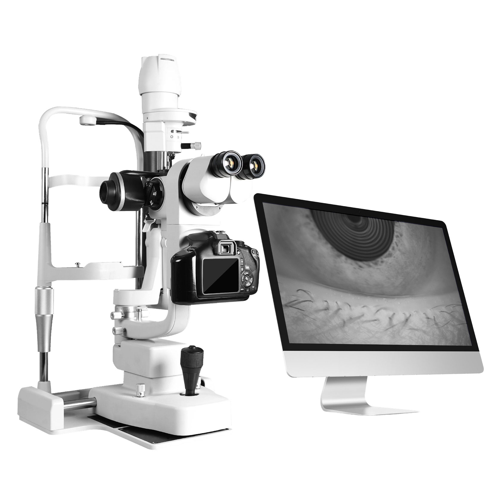
Click here to view the pdf catalogue
FEATURES
Optical microscopy combined with high-definition digital imaging to provide cell-level images.
Non-irritating Infrared light illumination to ensure the accuracy of tear film measurement.
One-stop inspection for anterior segment, operate easily and high-efficiency.
Optical resolution is 1900 lp/mm
Comprehensive Detection Of Ocular Surface Microenvironment
High-difinition imaging with infrared illumination, non-invasive, without irritation, ensure to get clearer images, to get real situation of tear secretion; Optional automatic calculation of TMH, operate easily and conveniently.
Specialized interference illumination and image system to real-time monitoring the lipid layer’s colors, flows, uniformity; Automatic calculation of lipid layer thickness with 1nm precision; Recording incomplete blinking times to assess blink quality and determine the dry eye due to tear dynamic abnormalities.
Infrared illumination, non-invasive, non-irritating, ensure smooth examination; High-definition video real-time monitoring of tear film changes; Automatic calculation of the first and mean break-up time, providing a more objective and accurate assessments.
Objectively assess the meibomian glands loss, access the dry eye of lipid abnormality, supporting clinicians in immediate management of meibomian gland conditions; Optional automatic calculate the proportion of meibomian gland loss.